
The Annual Meeting of the Radiological Society of North America (RSNA) revealed several new imaging technologies and imaging applications for pediatric patients, which provided an improved diagnostic accuracy, safety, patient-friendliness, and cost-effectiveness compared to current standards. Listed below are specific imaging applications and innovations that the Stanford pediatric imaging team presented. Further information can be obtained from the online program, through the cited abstract number.
Artificial intelligence (AI)
AI applications for medical imaging in general and pediatric imaging in particular played a major role at the RSNA conference. As different presenters outlined at the conference, AI can aid in generating medical images with reduced radiation exposure, substantially decreased acquisition times, and improved image quality. AI can help to streamline workflows and eliminate redundant tasks, AI can be used to develop quantitative metrics for quality assessment and qualitative improvement, and AI can support computer-assisted diagnoses.
In order to enable predictive and preventive personalized medicine, highly annotated databases will be needed to provide standards of reference for health and disease states. Dr. Curtis Langlotz, Derek Merk, and team described CNNs, which were trained on the RhodyNet database, and consisted of 285,565 radiographs to predict radiologist-assigned acuity scores for pneumonia diagnosis (SSC08-03). Dr. Matt Lungren presented results of the Stanford Tuberculosis AI trial (SPSI24B) and related challenges of AI applications to global health (RC416C). Dr. Greg Zaharchuk and team presented a new AI approach for accelerated whole body PET data acquisition (SSG09). Dr. Benjamin Frank, Youngho Seo, and team presented a deep learning model, which can predict order-of-magnitude five-year health care expenditure with reasonable performance (SSC08-09). Dr. Daniel Rubin presented a new approach to use out of distribution detection to fix AI models in medical imaging (SSE14-06). Nearly all AI models for medical imaging fail silently on input data dissimilar from the training data. Dr. Rubin and team trained AI models to produce confidence estimates along with their original task predictions. This can inform radiologists about the level of confidence of AI predictions.
Dr. Safwan Halbi explained the impact of big data on population health (IR42). To facilitate the development of AI algorithms for pediatric imaging applications, imaging data obtained at pediatric hospitals worldwide need to be collected in shared image registries to obtain normative data about the growing child, optimize and standardize imaging strategies and reporting guidelines across institutions, and eliminate repeated or comparative imaging studies. Dr. Abraham Verghese explained in his plenary lecture that AI clearly offers amazing insights into the interpretation of images without getting fatigued (PS10E). But AI will not replace radiologists. AI might increase the precision of some image interpretations, thereby leaving more time for radiologists to interact with physician colleagues and patients.
The Internet of Humans
The Internet of Things describes a system of interconnected computing devices and digital machines, which can transfer and exchange data over a network without requiring human interaction. The internet of humans (not things) describes a seamless connection of humans through transfer and exchange of human data. Our patients and their families want to participate in evaluations of their health status and they embrace developments of self-measuring devices. As Dr. Sanjiv Sam Gambhir explained in his plenary lecture (PS30B), radiologists as expert diagnosticians are uniquely poised to integrate the information of patient-driven health-monitoring devices into traditional diagnostic approaches. There are 4.6 billion mobile-phone subscriptions worldwide and there are between 1–2 billion people accessing the internet every day. Active input from self-measuring and wearable devices at the population and individual level will allow us to tackle key clinical questions regarding normal child development, patient compliance, therapy outcomes, and patient-perceived value. Successful development of electronic tools that can provide continuous rather than sporadic connections between patients and physicians will fundamentally change the way we approach human pathophysiology from detecting clinical symptoms to predicting diseases, initiation of preventive therapies, and maintaining human health.
Ultrasound Innovations
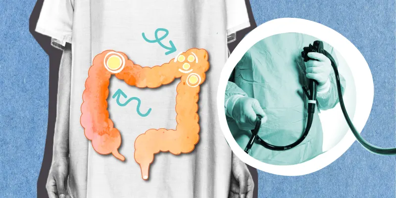
Drs. Jesse Sandberg, Erika Rubesova, and team discussed the value of microbubble-enhanced ultrasound for the diagnosis of inflammatory bowel disease (RC413-12). Members of the Stanford imaging team also discussed the POCUS initiative at Stanford: Our faculty believe POCUS will be an integral part of the practice of all family physicians in the next 5–10 years and are committed to training our residents in its use. To maximize resident experience and minimize logistical barriers, our program has made the commitment to put a personal POCUS device (Butterfly IQ) in the hands of every resident and every faculty physician. As of July 2019, we have provided devices to all second and third year residents and half of the faculty. At the RSNA industry exhibition, several vendors presented point-of-care ultrasound devices with ultrasound transducers, which can be connected to an iPhone (Butterfly IQ), Android phone (Philips Lumify) and/or tablet (e.g. Vscan, GE Healthcare). There were also new ultrasound technologies with wireless transducers, such as the SonoQue for the iPhone (KPI Healthcare) and the wireless Acuson Freestyle (Siemens), among others. These devices are relatively inexpensive and easy to acquire and operate, enabling the non-imaging expert to generate ultrasound images of the body with reasonable image quality for instant diagnoses in out-of-office settings. While early adopters include emergency physicians and family medicine physicians, possible future users might be patients with chronic diseases, who monitor their own condition at home. Radiologists will have to develop mechanisms for remote review of such point-of-care images, along with strategies for documented consultations and patient stratification to more advanced imaging tests.
Whole Body Cancer Staging
Several groups compared the diagnostic accuracy of whole body diffusion weighted MR images (DW MRI) and 18F-FDG positron emission tomography, with variable results, depending on tumor type, imaging protocol, and timing of the scan. Dr. Mary Ellen Koran and team compared unenhanced DWI with 18F-FDG PET and found overall improved performance 18F-FDG PET compared to DW MRI (RC613-03). Dr. Ashok Theruvath and team compared ferumoxytol-nanoparticle enhanced DWI with 18F-FDG PET and found significant agreement between treatment response assessments of pediatric lymphomas and sarcomas with the two imaging techniques (Α=0.91). Predictions of clinical response were not significantly different (p=0.58) using change in SUVmax (0.90; 95% CI: 0.81-0.97) and ADCmin (0.83; 95% CI: 0.55-0.99). However, in some patients, chemotherapy-induced changes in tumor metabolism preceded changes in proton diffusion (RC513-06). Dr. Anne Muehe presented interesting case studies of false positive findings on 18F-FDG PET/MR in pediatric oncology patients (PD172-ED-SUB8). All teams concluded that combined WB DWI and 18F-FDG PET provided the highest diagnostic accuracies.
Dr. Xuezhu Zhang from the University of California at Davis and colleagues (SSE23-06) presented results of total-body PET/CT studies with the Explorer. This new PET/CT scanner technology harbors a head-to-toe pet detector design with a 194 cm longitudinal field of view, providing state-of-the-art diagnostic PET images of the whole body with very small amounts of radiotracer activity (< 1 mCi), very short acquisition times (< 2 mins), and total-body pharmacokinetic studies with frame durations as short as 1 second.
Advanced Imaging Techniques and New Imaging Biomarkers
Dr. Lane Donnelly discussed tools for successful and sustainable quality improvement (RC613-09). Dr. Donald Frush discussed new approaches for CT dose monitoring (RC324). Dr. Vasanawala discussed 4D flow MR imaging techniques (RC512-05) and Drs. Zucker, Newman, Vasanawala, Barth, Fleischman and Chang presented applications for imaging of congenital pulmonary artery anomalies (PD130-ED-X). Several groups described the use of nanoparticles as alternatives to gadolinium chelates. Dr. Daldrup-Link presented an overview on iron oxide nanoparticle-enhanced MRI as a potential alternative to gadolinium chelate enhanced MRI (MSMI21C). Dr. Hossein Nejadnik described “one stop” labeling approaches of stem cells with nanoparticles for in vivo stem cell tracking with MRI (S505AB). Dr. Florian Siedek compared the value of ferumoxytol nanoparticle and gadolinium chelate-enhanced MRI for assessment of sarcomas in children and adolescents (RC513-05). Dr. Siedek’s study was recognized with an RSNA research trainee award.