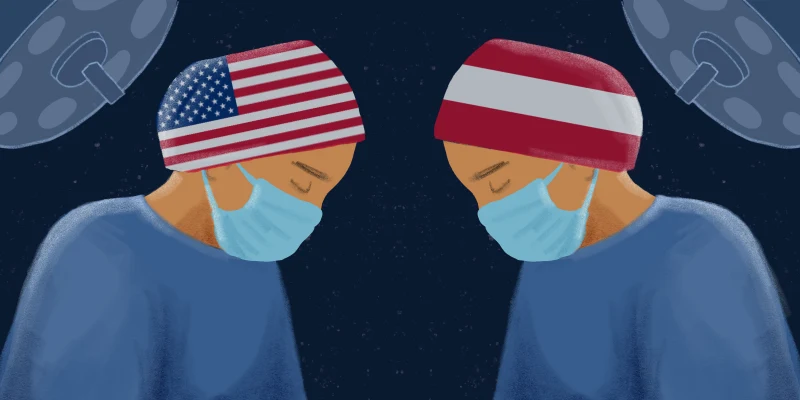
Gastroenterologists are among the many physicians experiencing burnout. They spend too much time in front of a computer clicking and typing out procedure reports, and not enough time with their patients. We believe artificial intelligence (AI) can solve this problem.
Imagine: While performing a colonoscopy, AI algorithms “observe” and record all the elements that need to go into that procedure note. If your report is generated reliably by AI, imagine how much more quality time you could have with your patient.
At the 2018 American College of Gastroenterology annual conference in Philadelphia, PA, our research team from the University of California, Irvine and Docbot was honored with 3 Presidential Poster Awards, a Category Award and a plenary session for research we have conducted using AI applications in colonoscopy and capsule endoscopy.
If you are a colonoscopist performing an AI-assisted colonoscopy, AI can reliably determine times of insertion and withdrawal, cecal intubation rate, prep quality, findings, locations, and tools used. Now, imagine that AI has documented your procedure, CPT codes, and quality measures. You are not burdened creating a report from memory later that day or evening and waiting for 1–2 weeks on a path report before “closing the loop”— you can instead review, expand, and sign at the point of care. AI can convert documentation time to quality time with your patients, all while reducing errors and costs associated with quality reporting and billing.
Is this a dream or soon-to-be reality?
Deep learning with convolutional neural networks is a form of AI that excels at image recognition, adept at characterizing images for the presence and characterization of any finding defined by the “trainer” on that image. A video is a sequence of images. As such, AI during an endoscopic procedure has the potential to aid in the detection of normal, landmarks, and disease states. AI learns from its trainers. Its accuracy can only be as good as its trainer and is limited only by the size and quality if its dataset.
Endoscopic technology has advanced tremendously, focused on image quality and field of view. We now have the ability to examine the gastrointestinal lumen with unprecedentedly high-definition (HD) clarity and field of view. Yet, among gastroenterologists using HD equipment, there remains significant variability in the detection of clinically relevant findings such as adenomas or flat sessile serrated polyps. Why is this? Location (e.g. cecum) and diagnosis of disease (e.g. precancerous polyps) require recognition and interpretation in real-time. This is a specialized ability dependent on multiple factors such as motivation, training, manual skills, and devices to clean and visualize the entire surface area. Beyond these are intrinsic abilities such as observer-dependent visual acuity and pattern recognition.
Imagine an expert gastroenterologist is standing alongside (as in fellowship training) and highlighting landmarks and areas of a potentially significant finding, while you drive the scope. The expert is doing this while at the same time teaching and training the endoscopist on how to heighten their own skills.
This is what AI can do if trained by experts.
A pertinent example is adenoma detection. Adenoma detection rate ranges between 7 and 54% among colonoscopists. The prevalence of adenomas is estimated to be > 50%. If a gastroenterologist misses one or more adenomas or sessile serrated polyps during colonoscopy, their patient has an increased risk for developing colon cancer before their next colonoscopy. Unfortunately, detection rates of precancerous polyps among colonoscopists range between 7–54% with a mean of ~30%. All colonoscopists should aspire to an ADR > 50%. Can AI help close this gap between ADR and actual prevalence for all colonoscopists?
Using a dataset of 8000+ HD images, we trained an AI algorithm to identify polyps in real-time during colonoscopy. We published our findings in Gastroenterology, showing 94% accuracy for polyp presence on images. In colonoscopy videos, AI-assisted expert colonoscopists detected 20% more polyps than they found when viewing the videos without AI assistance. Our next generation algorithm based on tens of thousands of polyp images has an accuracy of 99.5% and runs real time during colonoscopy. While these results are promising, it is too soon to know if AI can close the prevalence/detection gap in practice. This claim will require multiple unbiased randomized multicenter studies.
The potential for aiding polyp detection is only part of the story. At the plenary podium at ACG 2018, we showed that AI can differentiate precancerous polyps from serrated polyps with high accuracy, opening the possibility of automated adenoma detection rates, real-time colonoscopy interval recommendations, and (in the more distant future) adoption of “resect and discard” and “leave alone” strategies. Another one of our Presidential Posters demonstrated AI’s ability to define the presence and type of tools used during a colonoscopy.
Gastroenterologists are burdened on multiple fronts, and AI promises to automate the collection of data. AI may also assist in accurate billing codes. So, now imagine performing a colonoscopy with multiple AIs providing real-time feedback on prep quality, insertion/withdrawal times, cecal intubation, polyp detection, predicted size, morphology and pathology, techniques used to remove the polyp, and the presence of residual polyp tissue after polypectomy. Your report is essentially complete and mapped to appropriate CPT codes. All that is left that AI cannot yet fill in is the indication, last colonoscopy, and sedation used.
AI is the future. The bottom line is that it will reduce the time for documentation and maximize time with patients. The dream is no longer science fiction. It is rapidly approaching reality and can be applied across all areas of medicine. Wherever documentation is a burden or diagnosis is operator-dependent, AI can help us all be experts and escape from burnout, come home earlier, energized, and looking forward to working again tomorrow.
Dr. Jason B. Samarasena is an associate professor of medicine and the director of advanced endoscopic imaging at the H. H. Chao Comprehensive Digestive Disease Center at the University of California, Irvine.
Dr. William Karnes is a professor and the director of the High Risk Colorectal Cancer Program/Endoscopy Quality at the H. H. Chao Comprehensive Digestive Disease Center at the University of California, Irvine.
Dr. Karnes and Dr. Samarasena and are co-founders of Docbot Inc.