

Perhaps the most important aspect of a physician’s role is our diagnostic capabilities. Truly, if we cannot identify and diagnose a patient’s pathology, with reasonable accuracy, we cannot effectively treat them and may even cause greater harm.
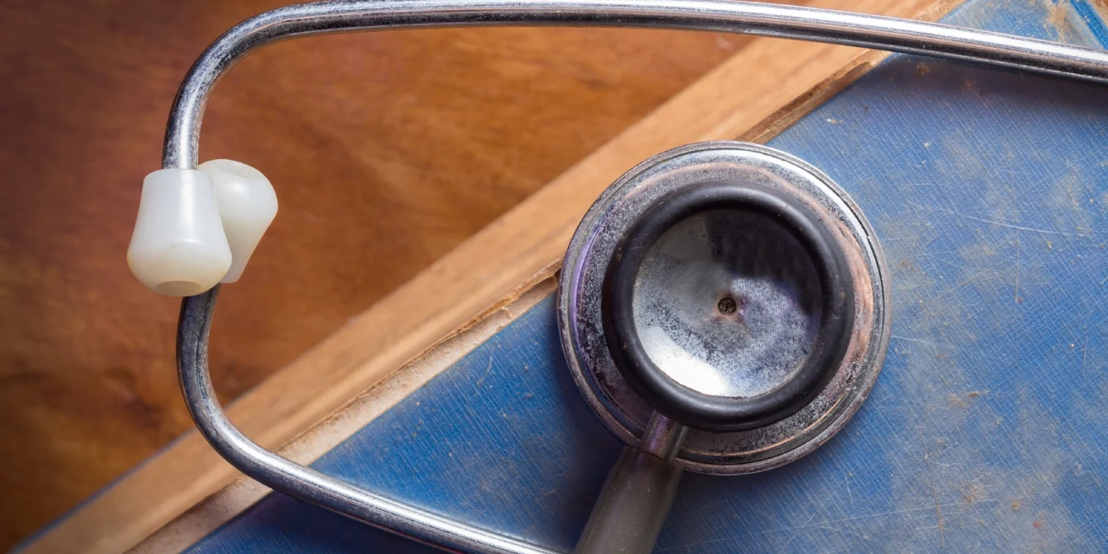
Let’s look back. The year is 1816 and you are a physician evaluating a patient with shortness of breath. The common practice of the time was direct auscultation by placement of the practitioner’s ear on the patient’s chest. At the time, many criticized this technique as providing little insight into the patient’s condition as well being out of normal social standards. Secondary to these pressures, that year, a French physician, Dr. Rene Laennec, invented the stethoscope, which dramatically improved the clarity of the auscultation exam. He spent the remainder of his career correlating how the stethoscope could detect various chest pathologies, and educating the utility of this technology on improving bedside assessment. The accumulation of his works resides in his text De l’Auscultation Médiate, which is one of the most widely translated medical texts in history.
Now, just over two-hundred years later, it is incredible to think that this technology has not been disrupted, especially when you compare the advances mankind has made in other areas. We have gone from the horse and carriage to cars that can practically drive themselves; from the notepad to the iPad, and can now communicate anywhere on the globe irrespective of language or distance. In health care as well, we have drastically altered our ability to monitor as well as treat our patients.
Yet despite all of our technologic advances, the stethoscope remains the primary tool used at the bedside for the physical exam. This is in spite of the widely known inaccuracies produced by the stethoscope guided physical exam. For example, studies have demonstrated dismal results regarding the ability of the stethoscope to detect common cardiac events with a reported accuracy of 20 to 40 percent. (1, 2) Even amongst cardiologists, the accuracy of the stethoscope is poor. (3)
Acceptance of this concept is key for us to innovate. One simply needs to ask themselves: how do you tell the difference between rhonchi or rales? What section of the lung did you hear diminished breath sounds? Or, where is that heart murmur emitting from and what pathology does it represent to realize the severe limitations of the stethoscope? Moreover, when you compare the scrutiny that we require all modern medical monitors to achieve for their accuracy (no one would use a pulse oximeter that had only a 40% percent accuracy rate), why do we not seek a similar level of accuracy when we discuss the tools we use for our physical exam? Fortunately, technologic advances have recently allowed for a new technology to be evaluated as an adjunct for our physical exam: point of care ultrasound (POCUS).
Similar to the evolution of computers, ultrasound machines have become much more portable (some are even pocket size) with functionality similar to “high-end” units used in imaging laboratories. New technologies even allow one’s smartphone to become an ultrasound device. This innovation allows the physician to have access to ultrasound imaging during their physical exam at the point of care.
The utility of POCUS has been demonstrated for nearly every type of physical exam including cardiac, pulmonary, neurologic, pulmonary, and abdominal. How often have you wondered: what is the ventricular function, how severe is that aortic stenosis, how bad is the COPD, is there a pneumothorax, what is the amount gastric volume, or what is the patient’s volume status? These are all questions that have proven to be answered “real-time” by POCUS.
Sadly, the utility for POCUS to facilitate acute care management has mostly been shown in emergency medicine and critical care specialties. (4) This has been viewed with some criticism given the fact that patients have the same comorbidities and acute care events in the perioperative setting. (5) Truly, from a patient care standpoint, why should the skillset for bedside evaluation of the patient change simply because they transition from the emergency room or intensive care unit to the perioperative setting?
Fortunately, the utility of POCUS to improve patient care in the hospital and ambulatory perioperative settings is starting to rapidly emerge. (6, 7) Interest in perioperative point of care ultrasound (P-POCUS) has been reported by anesthesiology trainees along with the integration of P-POCUS curriculums into residency training. (8) Moreover, the implementation of a perioperative POCUS service has also demonstrated a positive clinical impact. (8)
While it is encouraging to see the growth of P-POCUS there is much more that can be developed. Other specialties have developed formalized educational and certification pathways. (5) Emergency medicine has adopted POCUS training as a “core competency” for residency training. Currently, the certification and educational processes for the perioperative setting are limited, but the concept is growing.
A collaboration of academic programs including Loma Linda University Medical Center, UCI Medical Center, and UCLA Medical Center are working together to help address the need for education and training on this topic. Every year, more academic programs are developing training curriculums on P-POCUS.
One such curriculum, termed F.O.R.E.S.I.G.H.T. (Focused periOperative Risk Evaluation Sonography Involving Gastroabdominal Hemodynamic and Transthoracic ultrasound ) has been published as an effective strategy for education. This curriculum incorporates the topics of 1) Cardiac, 2) Pulmonary, 3) Hemodynamic, 4) Gastro-Abdominal 5) Airway, 6) Vascular access, and 7) Intracranial pressure assessment and highlights their relevance for the perioperative setting. To further P-POCUS education this curriculum is now online and is open access, under a Creative Commons license. Additionally, training workshops on this curriculum are now available as well.
Importantly, this is just one initiative to further the development of P-POCUS. Others include the Focus Assessed Transthoracic Echocardiography (FATE) protocol, which was developed by an anesthesiologist and is one of the most widely referenced POCUS examination protocols. Additional online resources available for education on P-POCUS include those from the Society of Critical Care Medicine and American Institute of Ultrasound. Finally, a recent review article highlighted the current CME training programs available on this topic. (5)
While these resources serve a key role, the impetus is on our specialty to develop structured guidelines, endorsed educational pathways, and certified credentialing processes to incorporate this new assessment tool into everyday practice. In 2016, a multi-subspecialty task force reviewed the current applications of P-POCUS and its training. (5) This group reported a “call to action” on this topic and emphasized the importance of our specialty’s societies to develop standards of training such that this modality is widely adopted amongst perioperative providers. (5)
Overall, while there is still much to be developed, the concept of P-POCUS has shown dramatic growth in recent years. Further innovation and integration of this tool have tremendous potential for improving perioperative care if we are willing to innovate. It is important for our specialty to accept how Point Of Care Ultrasound can positively impact patient care by providing real-time answers to many acute care events. To draw back to the works of Dr. Laennec, in 1834 (18 years after his innovation), the Times of London reported that the medical profession was unlikely ever to start using the stethoscope, “because its beneficial application requires much time and gives a good bit of trouble.” Clearly, Dr. Laennec efforts, through his education and texts, have proven this statement to be false. Now, however, we are at the same crossroads with the stethoscope and point of care ultrasound.
References
- Mangione S, Nieman LZ, Gracely E, Kaye D. The teaching and practice of cardiac auscultation during internal medicine and cardiology training. A nationwide survey. Ann Intern Med 1993;119:47–54.
2. Paauw DS, Wenrich MD, Curtis JR, Carline JD, Ramsey PG. Ability of primary care physicians to recognize physical findings associated with HIV infection. JAMA 1995;274:1380–2.
3. Kaul S. Is it Time to Replace Physical Examination with a Hand-Held Ultrasound Device? J Cardiovasc Echogr 2014;24:97–102.
4. Arntfield RT, Millington SJ. Point of care cardiac ultrasound applications in the emergency department and intensive care unit — a review. Curr Cardiol Rev 2012;8:98–108.
5. Mahmood F, Matyal R, Skubas N, Montealegre-Gallegos M, Swaminathan M, Denault A, Sniecinski R, Mitchell JD, Taylor M, Haskins S, Shahul S, Oren-Grinberg A, Wouters P, Shook D, Reeves ST. Perioperative Ultrasound Training in Anesthesiology: A Call to Action. Anesth Analg 2016;122:1794–804.
6. Khoche S, Ramsingh D, Maus T. The Year in Perioperative Echocardiography: Selected Highlights From 2016. J Cardiothorac Vasc Anesth 2017;31:1554–61.
7. Deshpande R, Ramsingh D. Perioperative point of care ultrasound in ambulatory anesthesia: thinking beyond nerve blocks. Curr Opin Anaesthesiol 2017;30:663–9.
8. Ramsingh D, Rinehart J, Kain Z, Strom S, Canales C, Alexander B, Capatina A, Ma M, Le KV, Cannesson M. Impact assessment of perioperative point-of-care ultrasound training on anesthesiology residents. Anesthesiology 2015;123:670–82.