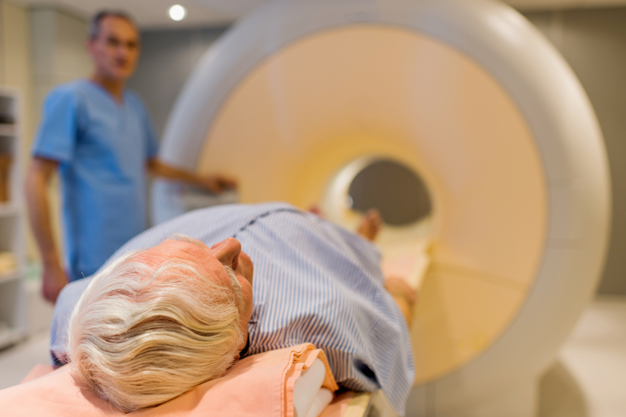
As a general urologist, it is becoming increasingly important to understand and interpret the nuances of prostate MRI. With the results of the PROMIS and PRECISION trials presented at the recent 2019 American Urological Association Annual Meeting, it is safe to assume that prostate MRI is here to stay for the foreseeable future.
The hands-on courses at the meeting this year provided an excellent opportunity for the novice interpreter of prostate MRI to gain a significant amount of expertise in understanding and interpreting these complex imaging studies.
The first take-home message is understanding why prostate MRI can distinguish high-grade prostate cancer from benign prostate tissue and indolent prostate cancer. This capability of MRI is based upon the water content of the tissues. Due to the higher water content of benign prostate tissue and indolent prostate cancer, these tissues demonstrate a different signal intensity as compared to high-grade prostate cancer, which has less water density.
Once the urologist understands the basic concepts of MRI for prostate cancer detection, they must consider the technical factors in how the MRI is performed. There is no universally accepted protocol for performance of prostate MRI. Individual institutions have developed their own techniques to maximize the quality of the imaging. Important factors to consider for maximizing quality include eliminating air from the rectum, which can be done with an enema and/or encouraging the patient to pass flatus immediately prior to imaging. Further improvement in the quality may be achieved with the implementation of an endorectal coil, although this can be uncomfortable for patients. The magnet size can also be important in maximizing imaging quality, and henceforth many centers utilize a 3T magnet rather than a 1.5T. Ultimately, each institution must develop its own protocol to achieve quality images.
Expertise in interpreting prostate MRI is critical for accurate diagnostic performance. When viewing the MRI, one must review three key sequences. It is a pitfall to evaluate any single sequence individually. These three sequences include the T2, the diffusion sequences (diffusion-weighted imaging (DWI), and apparent diffusion coefficient (ADC), and the dynamic contrast enhancement sequence.
Let’s start with the T2 weighted images. On these images, tissues with high water content will appear bright white. Conversely, prostate cancer will appear dark on T2 weighted images. Again, this is due to the high cell density of prostate cancer. If a concerning dark area is seen on T2, this area should be examined on the DWI and ADC images.
The DWI and ADC are similar in that they reflect the diffusion of water within the tissue. It should be noted that the DWI are used to create the ADC images. The ADC images are essentially a composite of the DWI images at several different B values. The diffusion-weighted images may be viewed at various B-values, which should be set to at least 1400 in order to accurately identify lesions concerning for prostate cancer.
The bottom line for DWI and ADC: Prostate cancer is bright white on DWI and dark on ADC. (Easy way to remember this: DWI has “WI” that is short for white; ADC has “DC” that is short for dark.
The third sequence to consider is the dynamic contrast enhancement (DCE) sequence. These images should be viewed as a cinematic view over time as the prostate takes up the contrast. A key concept for interpreting these images is the understanding that prostate cancer will demonstrate rapid uptake and rapid wash out of contrast.
Using the information gathered from the T2, diffusion (DWI and ADC), and the DCE sequences, the urologist should be able to identify a significant prostate cancer as dark on T2, bright white on DWI, dark on ADC, and avidly enhance on DCE.
After interpreting a prostate MRI, it is important for the radiologist to create a report that is standardized. This should involve the utilization of PIRADs v2.1 in order to stratify the risk of prostate cancer into five categories, ranging from 1 (very low risk) to 5 (very high risk). It is also important to understand the limitations and pitfalls of prostate MRI, for example, granulomatous prostatitis will mimic a PIRADs 5 prostate cancer. It should also be noted that the PIRADs scoring system should not be used in post-radiotherapy cases.
In addition to providing an excellent review on the interpretation of prostate MRI, this year‘s AUA hands-on course for MRI-US fusion included an introduction to three separate fusion prostate biopsy systems. In no particular order, the three systems were Koelis, UroNav, and Artemis.
The Koelis system is based upon a mechanism within the tip of the US probe, which repeatedly rescans/sweeps the prostate prior to and following each firing of the biopsy needle. UroNav’s system is based upon an electromagnetic field generator that tracks a marker secured to the ultrasound probe. The Artemis device utilizes a robotic arm to hold the ultrasound probe. All three systems each have pros and cons, so I would recommend urologists have trials each device before committing to any one platform.
The role of MRI in prostate cancer will only continue to expand. MRI serves as a powerful tool that can guide decisions for the management of prostate cancer and reduce the detection of indolent prostate cancers.
Richard Knight, MD is a urologist at the 48th Medical Group, RAF Lakenheath, UK.


