

Throughout the particularly stressful time of residency and fellowship interviews, there is probably no greater bright spot than having the opportunity to interact with your future colleagues and contemporaries on the “interview trail.” So it was for me, in 1996, as a relatively green ophthalmology resident, as I interviewed for a fellowship in corneal and refractive surgery. Throughout my travels across the country that fall, I came across a particularly bright fellow, David Huang, MD, PhD. As we began talking, I remember him discussing research he had been involved with in college and during his residency, about a new technology that he co-founded called optical coherence tomography, or OCT. At the time, I knew very little about it, and cared probably even less. After all, I had bigger fish to fry. Fast forward 24 years, and the OCT is probably the one piece of diagnostic equipment that I absolutely cannot practice ophthalmology without.
I explain to patients that OCT is like a “CAT Scan” for the eye. It allows us to take virtual “slices” and put together images that can analyze various layers throughout the eye. The most commonly used benefits of OCT lie in diagnosing subtle macular pathology and its utility in assessing the retinal nerve fiber layer for early signs of damage in patients with open angle glaucoma. However, today, we are using OCT in ways that, just maybe, even Dr. Huang never imagined. (But if I know, David, he probably did.)
Many cataract surgeons are using femtosecond lasers in their craft, and these lasers require precision placement of the pulses within the lens in order to create the capsulotomy, and lens fragmentation. In addition, the corneal anatomy is important when creating incisions for phacoemulsification and correction of astigmatism. This is all made possible by OCT: all laser platforms utilize OCT in order to create a registration image of the eye so that accurate laser delivery can be administered.
OCT has also been introduced into the operating microscopes that we use for corneal and retinal surgery. In cases of retinal membrane peeling, Descemet’s Membrane Endothelilal Keratoplasty (DMEK), Deep Anterior Lamellar Keratoplasty (DALK), among others, having the ability to visualize the various layers of the eye as well as the behavior of the implanted tissue may allow these procedures to be performed with much greater success.
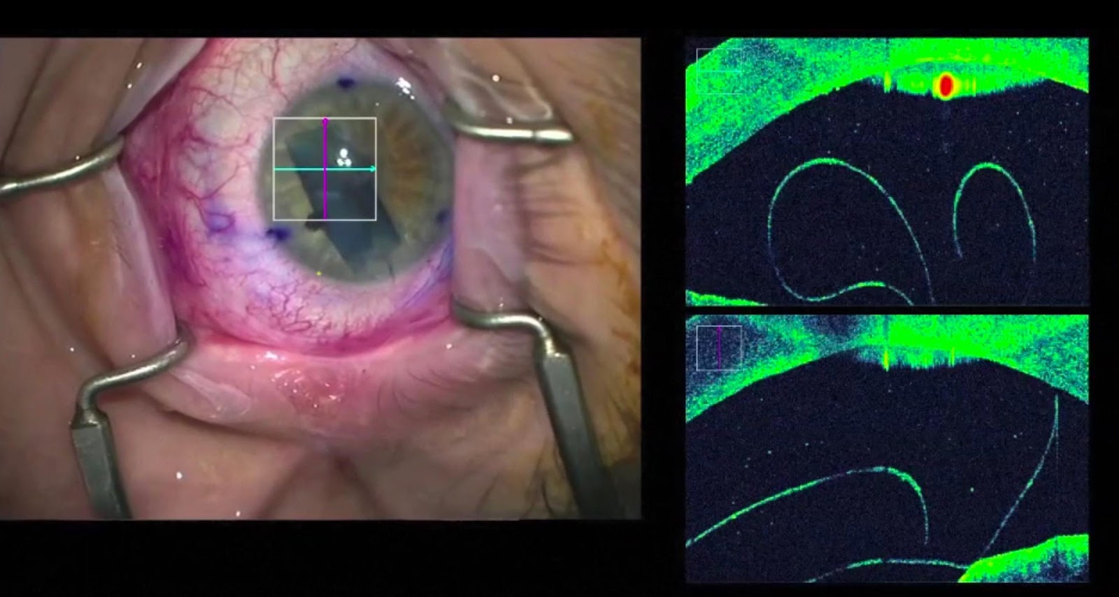
Fig. 1. Real-time OCT showing the configuration of the DMEK graft in the anterior chamber.
However, in my practice, the OCT has proven indispensable in two major areas. As an early proponent of DSAEK since 2007 and DMEK since 2013, I began using OCT to help assess immediate postoperative graft adhesion, and to this day, it remains an invaluable tool in planning for optimal management of these grafts. Especially in cases of postoperative corneal edema, OCT provides immediate, non-invasive feedback as to graft position and can predict the likelihood of graft success with or without further intervention.
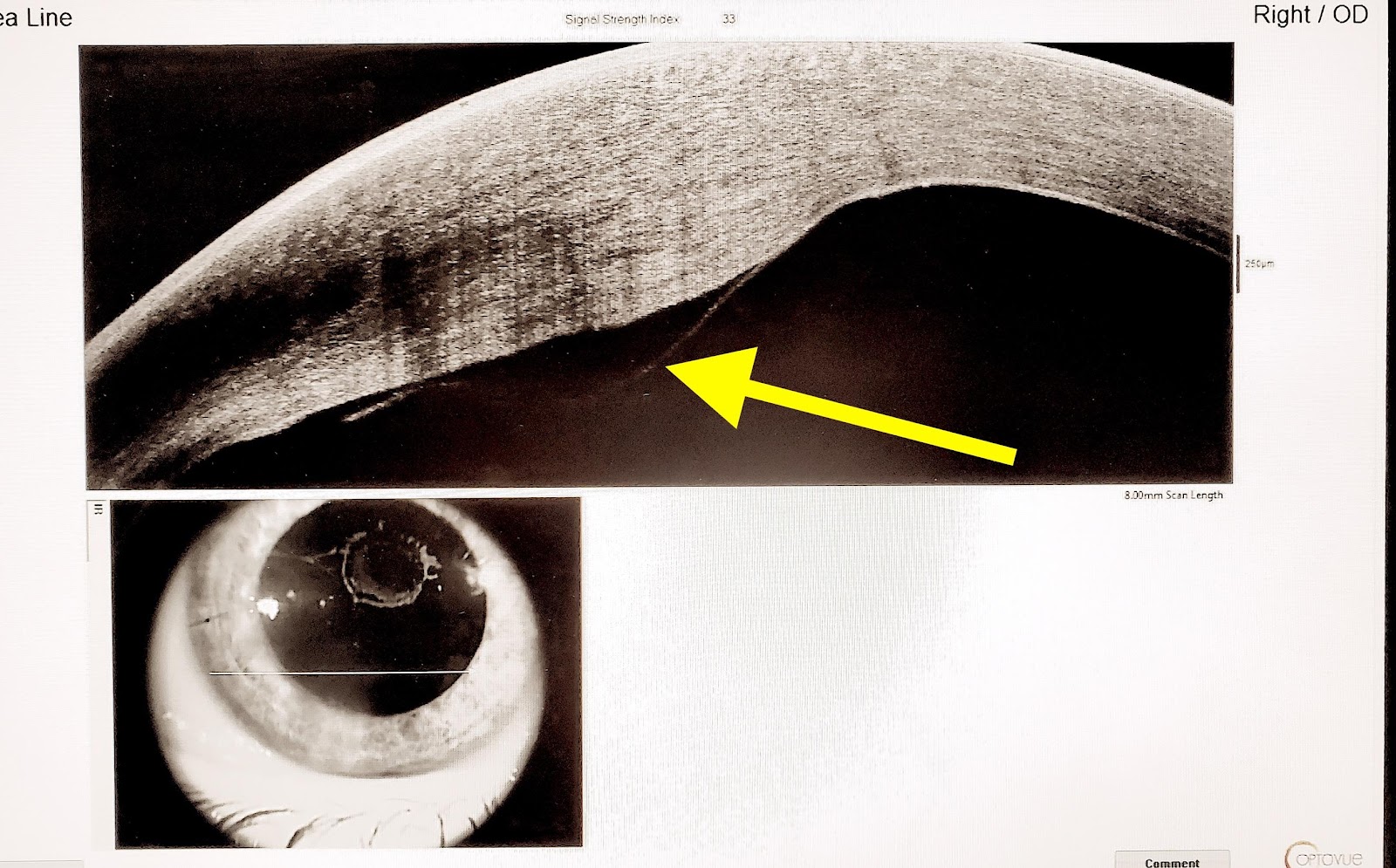
Fig. 2. Anterior Segment OCT showing DMEK graft detachment
A second, but possibly slightly lesser known, benefit of OCT lies in the ability to accurately assess the true corneal power. It is commonly accepted that following LASIK surgery, intraocular lens (IOL) calculations may be erroneous, unless specific factors are taken into consideration. There are many formulas that can correct the commonly used anterior topographic measurements, but in my opinion, the OCT can quickly and accurately provide a calculation that will help to derive the correct IOL power that needs to be implanted. In fact, the Hill-Wang-Koch online IOL power calculator (through the American Society of Cataract and Refractive Surgery) has incorporated OCT power measurements, thereby validating OCT as a reliable source for pre-operative information.
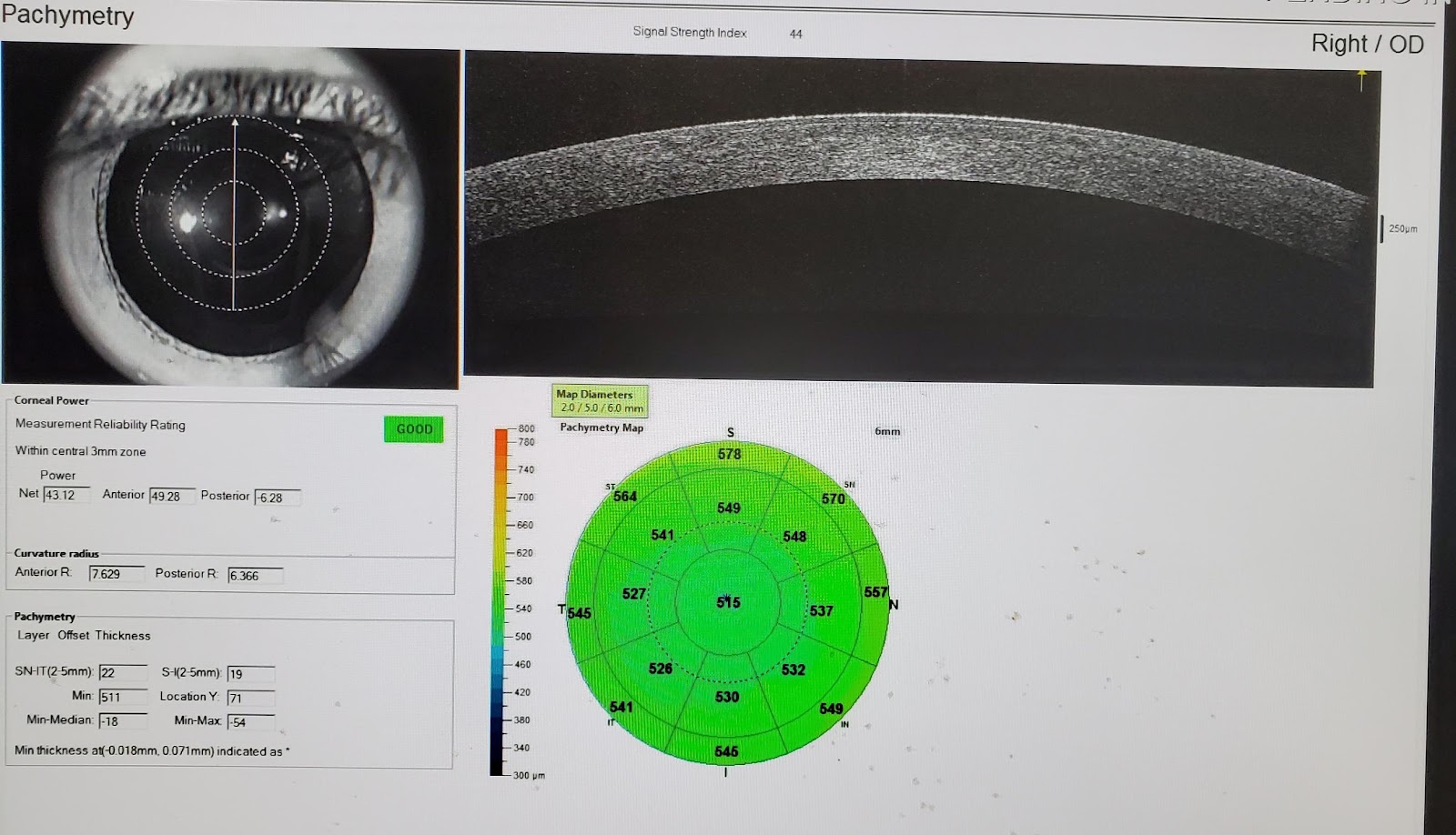
Fig. 3. OCT measurement of post-LASIK anterior and posterior corneal power
I’ve been fortunate in my nearly quarter century in this field to have to come across many innovative and exceptional surgeons, some of whom have been mentors and colleagues, and others who have been my residents and fellows. Among my fondest memories in ophthalmology will always be meeting David decades ago on the interview trail, and having had the opportunity to truly glimpse the future of ophthalmology.