
The ongoing COVID-19 pandemic has led to a sudden and exponential surge in workload for health care systems across the globe. Early identification of COVID-19 has also proved challenging, with numerous reports of individuals initially testing negative for the coronavirus (SARS-CoV-2) only to test positive on secondary testing.
The growing demand for more rapid and efficient triage of cases has spurred clinicians to explore new avenues for COVID-19 management more urgently than they would have under normal circumstances. Such efforts have led to the development of artificial intelligence (AI) algorithms primed to offer a helping hand to clinicians on the front lines of COVID-19 care.
In an interview with Doximity, physicians and AI luminaries discussed how AI has already begun to bolster the early detection of COVID-19 at their institutions, as well as the ways in which its role is likely to evolve in the days to come.
A Physician’s "Second Eye"
“‘Ten years of progress in a week’ — that’s the saying that’s been going around,” said Dr. Albert Hsiao, associate professor of radiology at the University of California, San Diego School of Medicine. “The coronavirus pandemic has forced the health care industry to greatly accelerate its pace of change — to look at new solutions that have been on the fringe and bring them toward the mainstream.”
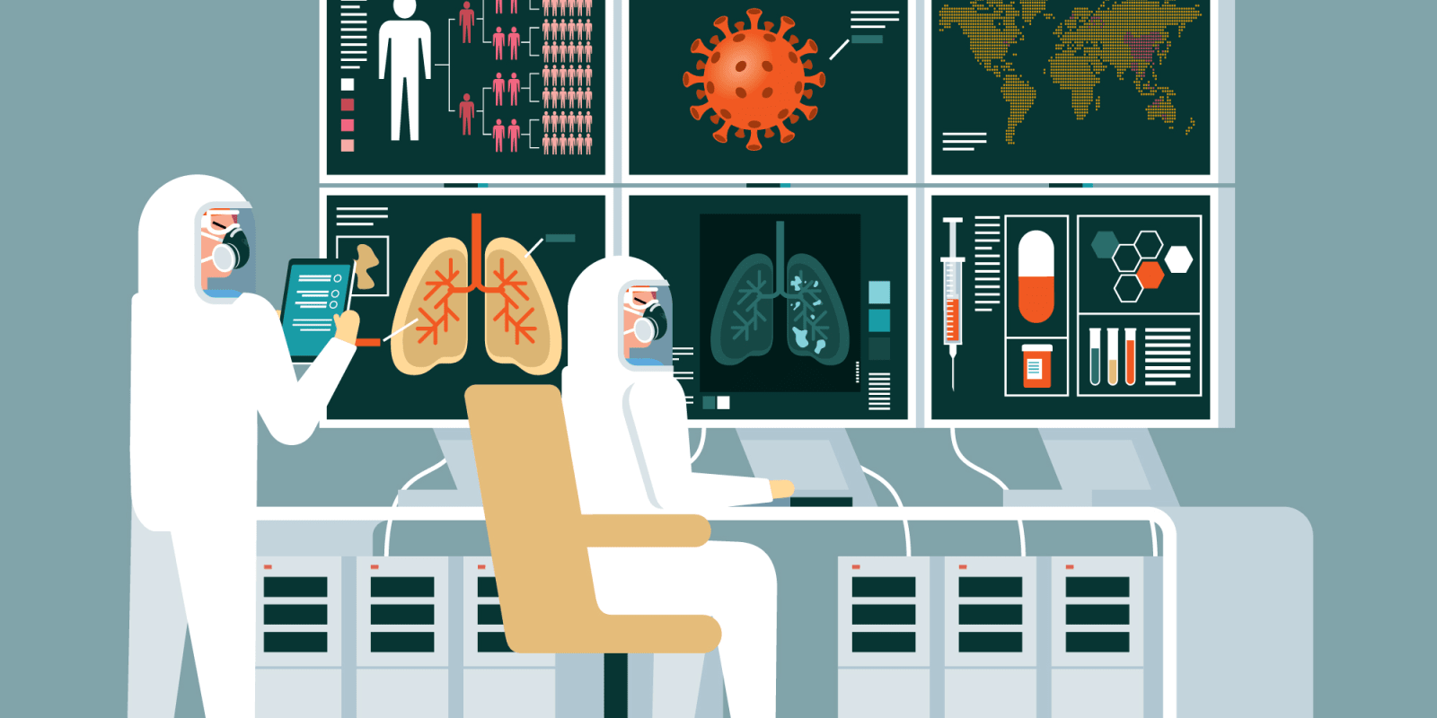
For their part, Dr. Hsiao and physician colleagues at UC San Diego Health have been exploring the use of AI as a tool to help physicians identify COVID-19 on imaging exams as quickly and accurately as possible. They have made their deep-learning algorithm, capable of automatically detecting pneumonia on X-ray images, available to clinicians at their medical center.
Pneumonia is one of the most common reasons leading to hospitalization in patients with COVID-19. By facilitating pneumonia diagnosis, the researchers’ AI algorithm has helped improve the diagnostic abilities of physicians at the point of care. It has acted as a kind of “second eye” to strained or less experienced physicians — highlighting subtle abnormalities and tracking disease progression in moderate to severe cases.
In a study recently published in the Journal of Thoracic Imaging, the researchers detailed the inner workings of their AI algorithm. They trained it on 22,000 annotated X-ray images to ensure it could accurately calculate the probability of pneumonia for each pixel of an X-ray image. The results are overlaid onto the original image with a color scheme that allows clinicians to distinguish between varying degrees of disease severity.
The researchers applied their AI algorithm to the X-ray images of COVID-19 patients treated at various hospitals in China and the U.S. For the five cases they analyzed in depth, the researchers found that the probability maps had correctly identified regions of the lung affected by pneumonia at every stage of disease severity. The algorithm maintained its effectiveness despite examining images sourced from a variety of X-ray machines at several different medical centers, underscoring its generalizability across institutions.
Scaling Up
In a matter of weeks, the pandemic catalyzed the integration of Dr. Hsiao's AI algorithm into the clinical diagnostic workflow at UC San Diego Health. Before COVID-19, it had been sidelined for more than a year and a half. Radiologists and physicians from other specialties have since applied the algorithm to well over 2,000 X-ray images.
A number of these X-ray images unexpectedly originated from patients who presented to the ED for reasons unrelated to COVID-19. If not for the AI algorithm flagging these patients’ images for review, their COVID-19 diagnosis would have been delayed, if not missed entirely.
“The galvanization of resources, not only to accept this AI technology but actually push it and get it to the point where it doesn’t cost the provider any extra time to consume, has been remarkable,” said radiology resident Dr. Brian Hurt, who developed the latest iteration of the group’s AI algorithm along with Dr. Hsiao.
What’s more, the researchers are continually improving upon their AI model as they get access to increasingly more COVID-19 imaging data and receive feedback from referring physicians.
“We have access to thousands of images and predictions at this point,” Dr. Hurt said. “This level of engagement will allow us to continue to scale up the AI algorithm and improve its skill level from a low level to a midlevel radiology resident. It’s been very exciting from that standpoint.”
"The next question is whether AI will be able to predict if patients’ initial symptoms will progress into more severe symptoms associated with COVID-19 or gradually resolve on their own," Dr. Hsiao added.
Looking ahead, the physicians are exploring avenues through which they can extend their AI algorithm to other institutions, enabling the community at large to better evaluate its effectiveness and boost its value for COVID-19 management. Their AI algorithm is accessible online for any clinician interested in appraising it using images acquired at any medical center.
“One of the major challenges is that it’s hard for physicians and health care systems to have confidence that AI algorithms will perform well in their clinical environment,” Dr. Hsiao said. “Perhaps the best way for folks to figure out whether an AI algorithm is valuable is to test it out themselves.”
Long-term Vision
Over on the East Coast, Dr. Eliot Siegel, Vice Chair of diagnostic radiology at the University of Maryland School of Medicine, has been working on a slightly different AI algorithm as part of a joint effort with Hayit Greenspan, PhD, head of the Medical Image Processing and Analysis Lab at Tel Aviv University and chief scientist of AI software company RADLogics.
The team of physicians and medical imaging experts has developed a deep-learning algorithm with the capacity to detect COVID-19 on CT scans. Beyond simply confirming the presence of COVID-19, the algorithm automatically produces a report containing volumetric measurements of ground glass opacities and other common imaging findings associated with the coronavirus and then displays the information as a 2D heat map as well as a full 3D visualization of the lung. Finally, the AI algorithm computes a “CoronaScore” that quantifies disease severity and progression.
They demonstrated the AI algorithm’s ability to distinguish between COVID-19 and non-COVID-19 cases with near-perfect accuracy (achieving an area under the curve of 0.996) in an exploratory study uploaded to arXiv.org.
Towards the end of January 2020, Dr. Greenspan and colleagues at RADlogics noticed a sudden uptick in China's use of her team’s AI algorithm. (Years prior to the outbreak, her team gave Chinese hospitals an early version of the AI algorithm that could detect and track common abnormalities on chest CT scans.)
As the prevalence of COVID-19 began to explode internationally, the researchers recognized the potential benefits of adapting their existing AI algorithm to detect the disease and thereby support physicians grappling with its diagnosis.
“What’s fascinating to me was the potential to be able to take AI software, modify one or two programs, and make it clinically useful in a really timely fashion, much in the way a virus might evolve over time,” Dr. Siegel said. “It only took about 10 days to accomplish this goal. This has important, positive implications for dealing with future pandemics.”
An Ideal Tool
Though still in a trial phase, the team’s AI algorithm has been integrated into the workflow of various health care networks in China, and is being deployed in Italy, Russia, and Brazil, among other sites. The researchers also are actively pursuing emergency use authorization from the U.S. FDA.
“It’s interesting to see the different approaches to COVID-19 detection and diagnosis all over the world,” Dr. Greenspan said. Currently, CT is considered an official method for diagnosing COVID-19 in certain regions of China, and is widely used in the Netherlands and several other European countries. In the U.S., however, chest imaging is not accepted as a first-line tool for COVID-19 management, though the RSNA and other societies recently published a consensus statement validating chest imaging to triage patients who present with moderate to severe clinical features or worsening status.
To that end, the researchers have made a move towards tracking the longer term consequences of the disease, which will prove critical once the first wave of cases subsides. They believe AI could ultimately help quantify disease burden, trace disease recovery, and reinforce differential diagnosis.
“We’re amassing a tremendous amount of data at this point — including from images, laboratory findings and clinical symptoms — and these multiparametric data are going to help us understand complications of the disease following the surge,” Dr. Siegel said.
“There’s been a lot of change in the medical imaging community and we feel AI can really make a difference,” Dr. Greenspan said. “We’re still not all the way there, but we are now reaching the point where we can start helping physicians, especially radiologists, save time, increase accuracy, and be an ideal supporting tool.”
To what extent do you believe AI could help clinicians manage the coronavirus pandemic?
Click here to see more perspectives on COVID-19 from the Doximity network.
Click here for up-to-date news about COVID-19 on Doximity.